|
Home
|
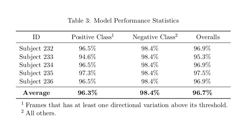
We employed a convolutional neural network (CNN) model for our classification task. Our model was able to achieve 98.3% and 96.4%accuracy on the positive (i.e., with-motion) and negative (i.e., without-motion) classes respectively. Table 3 presents the model’s performance on each individual subject.
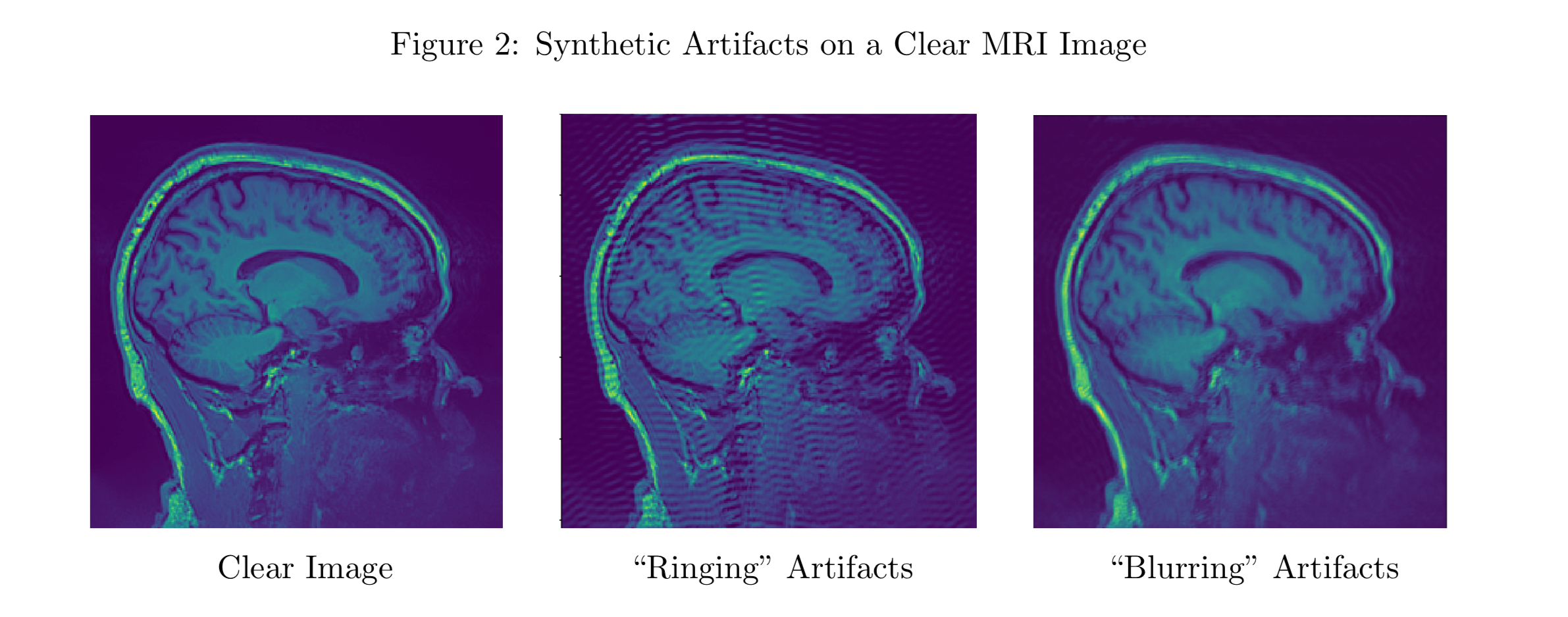
Code repository: https://github.com/hyuan9310/MRI_image.git Software Package: Coming Soon Reduce Image Artifacts Induced by In-scanner MotionsAlthough Deep learning has attracted a great amount of interest in recent years and has been widely explored in many medical applications, few studies exist in the area of correcting motion artifacts of MRI scans. The difficulty is two folds: 1) MRI images are obtained through a complex electronic and mechanical process using Fourier and inverse- Fourier transforms. Thus, the resulting motion artifacts (e.g., blurring) can’t be effectively modeled using traditional techniques such as convolution kernels. 2) The success of a deep learning model rests on the availability of a large quantity of training data. It is simply infeasible both financially and labor-wise to collect a huge amount of MRI images corrupted with motion artifacts.In our approach, we address the above two challenges by directly models the K-space representations of an MRI scan and generate sythetic artifacts that resemble the "rings" and blurs in the real motion corrupted scans. Figure 2 illustated the sythetics artifacts generated by our software:
We evaluated the performance of our model using a real-world dataset from NYU Langone's Comprehensive Epilepsy Center. The dataset consists of whole brain T1-weighted MRI scans obtained in six individuals imaged with both (i) deliberate head motion carried out during MRI acquisition and (ii) motion-free acquisitions. We further evaluated of our approach by applying the developed model to 55 MRI scans from the multi-center Autism Brain Imaging Data Exchange initiative. Figure 5 presents a sample of our experimental results. Code repository: https://github.com/yijun2011/tzo Software Package: Coming Soon |